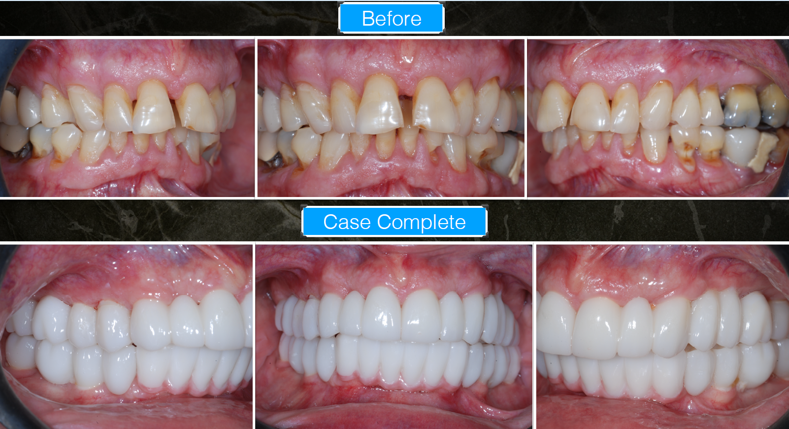
The case documents comprehensive rehabilitation of a patient with severe restorative, periodontal, and esthetic compromise requiring full mouth reconstruction. Initial facial and intraoral photographs demonstrated:
- Multiple failing restorations
- Missing and fractured teeth
- Malocclusion and uneven occlusal plane
- Severe anterior esthetic compromise
- Generalized wear and functional deterioration
Preoperative panoramic radiographs and 20 × 20 CT scans demonstrated extensive restorative history and compromised dentition requiring implant-supported rehabilitation.
Detailed clinical images revealed significant maxillary anterior tooth loss and fracture with compromised gingival architecture. The surgical phase involved minimally invasive extraction and placement of one-piece zirconia implants in the anterior maxilla.
Three-day postoperative photographs demonstrated rapid soft tissue healing and successful integration of immediate provisional restorations. The restorative workflow then progressed to:
- Initial final posterior restorations
- Anterior provisional restorations
- Occlusal rehabilitation
- Full arch esthetic refinement
Final “Case Complete” clinical photographs showed:
- Harmonized occlusion
- Improved facial esthetics
- Stable gingival contours
- Symmetric anterior restorations
- Full arch functional and esthetic rehabilitation
Final radiographic comparisons between preoperative and postoperative imaging demonstrated successful implant placement and full mouth restorative reconstruction.