CLINICAL FINDINGS
- Bilateral oro-antral fistulas
- Large posterior maxillary defects
- Scarred wound margins from previous surgeries
- Deficient posterior maxillary alveolar bone
- Peri-implant defects and soft-tissue compromise
- Deep peri-implant probing depths with suppuration in peri-implantitis cases
- CT scans demonstrated:
- Large bilateral posterior maxillary osseous defects
- Loss of sinus floor cortical plate
- Severe posterior maxillary bone loss
- Sinus communication defects
- Peri-implant crestal bone loss around tilted implants
- Sinus penetration and opacification in chronic rhinosinusitis cases
- Complications associated with:
- All-on-4 treatment
- Excessive bone reduction
- Poor wound closure
- Sinus membrane perforation
- Implant malposition
Importance of:
- Adequate keratinized tissue
- Proper implant depth and angulation
- Biofilm removal in peri-implantitis
- Proper sinus management
- CBCT-guided treatment planning
Advanced reconstructive techniques:
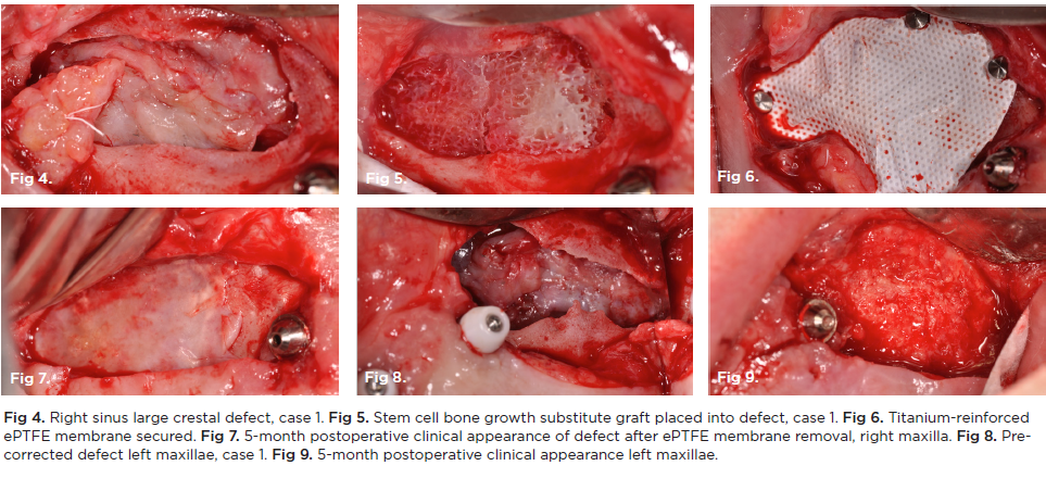
- Stem-cell enriched grafting
- Titanium-reinforced membranes
- PRP/A-PRF utilization
- Soft-tissue flap advancement
- MEMM (Modified Endoscopic Medial Maxillectomy)
Zygomatic implant complications:
- Chronic rhinosinusitis
- Implant sinus penetration
- Foreign-body reaction
- Biofilm colonization
Reported outcomes:
- Successful closure of all oro-antral defects
- 89.1% implant success in regenerated posterior maxilla
- Significant postoperative sinus improvement
Treatment Planning
TOPIC E - ALL ON X ALL ON 4 COMPLICATIONS
Persistent oro-antral communications after implant treatment Chronic sinus complications Failed prior surgical closure attempts Posterior maxillary bone loss and discomfort
.png)